
This blog comes to you at the conclusion of the year’s testing and evaluation. There are things we have noted and used in the lab. Then we break it down into the very best for the portable or point of care market and the console systems. The very best did not necessarily mean very expensive either. There are options here for all budgets.
2D imaging is the basis for all the diagnostic judgments in al specialties. In vascular the anterior borders and plaque detection is just as important as the posterior borders of the vessels. This is the prerequisite for the IMT (Intima Media Thickness) and plaque analysis features of the machines. Accurate plaque border and content detection is critical to the success of the ultrasound exam.
The issues in vascular ultrasound imaging are many. The machines need to have the ability to image very low flow and very high flow. Therefore the machines must be able to have a wall filter that has a wide enough variance to adjust for these extremes both in arterial and venous structures. The machine must have measurements that are customizable to allow for protocol specific vessels to be measured. The other alternative is a workaround/relabel to make sure that each segment of each vessel is annotated properly and then goes to the ultrasound report. If your machine does not have this capability then you need to reconsider your purchase.
Measurements need to have a variety of ways to make those measurements. For arterial measurements the most common measurements are the Peak Systolic Velocity and the End Diastolic velocity. There are various ways to perform those measurements. You can select just PS and Ed which are point and drag. You can do a manual trace from the start of the cycle to the end of end diastole. Then there is the auto measurement, which is not on all ultrasound machines. For this to work well the sonographer needs to have a great angle of insonation (less than 60 degrees) and the pulsed wave Doppler gain set correctly. The closer the angle is to 0 the more accurate your numbers will be. Automated measurements save much time and measures more than one cardiac cycle.
For extremely high velocities your machine may need continuous wave Doppler to get the most accurate peak velocity. Again, before you buy an ultrasound machine please consider this issue because not all ultrasound machines have CW options or a CW module in the machine at the time of purchase. Sometimes people forget that with CW the positioning of the entire CW line has to be only seen in the vessel you are examining. Continuous wave samples along the entire cursor line not just inside the sample volume. So if you hit a vessel anterior to where the vessel is you are interrogating then the Doppler tracing will show you both waveforms. This is how your accuracy will be adversely affected.
Then we need to mention color Flow Doppler. Again the wall filter has to be set properly and that may vary from patient to patient and vessel to vessel. The Priority has to be set correctly as well as the gain. It is important to note that the color box must be angled correctly to give you the best information. The scale or the number at the top and bottom of your color bar (Nyquist limit) must be set correctly as well. Most of these things need to have the flexibility and variance range to allow for all the changes the sonographer will make. Not all machines are flexible enough to do this. All these features hold true for power Doppler for very low flow imaging.
The best console systems that have all these previously mentioned benefits are the Mindray DC88, DC 70, the GE Logiq E10 and the Philips Affinity series. These machines have customization for measurements. The Color and Power Doppler are gorgeous. The GE has Auto Doppler Assistant that allows for faster Doppler adjustments. The Philips has a 3D linear probe that images the plaque well.
The best portable vascular ultrasound machines are the Mindray M8 and the Philips CX50. The Mindray Z60 and the TE5 have many of the above listed features but you cannot customize the measurements. In vascular the measurement customization is very important. Image quality directly affects disease diagnosis and is a critical piece of the exam.
This blog will allow you to judge which machine is best for your needs and budget. The machine families of Mindray, GE and Philips have many products for your needs as well. If you have any questions please call Ultra Select Medical at 843-566-1020.
This is the Philips Affinity 50G. This is the right CCA in #D volume rendered image. The light blue is the plaque and shows up beautifully.
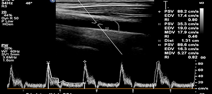
This is a Philips Affinity 50G showing the different ways measurements can be made. This first is an auto measurement, the second cycle is the standard two point and the last cardiac cycle is a manual trace.
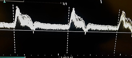
This is an example of an auto measure for three cardiac cycles. This was performed on a Mindray MX7 and there is no need to place the beginning and end cursors. It autodetects the beginning of systole as seen by the vertical dotted lines. You can see where error would be introduced if the Doppler gain was too high. Then Doppler angle is 45 degrees even though you cannot see it in this image.
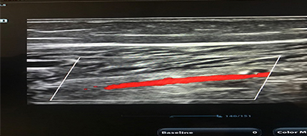
This is an example of low color priority (38) in the posterior tibial artery. This will not be fixed by increasing the gain or decreasing the scale.
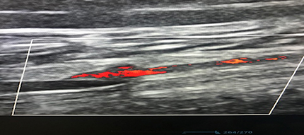
This is the same vessel as the one on above. This is 100% color priority. Very good color fill without bleeding into the soft tissue

Categories
The Very Best Vascular Ultrasound Machines