
There has been an explosion of new technologies in our field over the last couple of years. Some of the things we will discuss are not new but very useful. Based on our customer’s feedback, discussions with manufacturers and academic articles we feel that these are the ones that will give you added clinical value. I have to admit that it was a hard choice. We limited our blog to the best five.
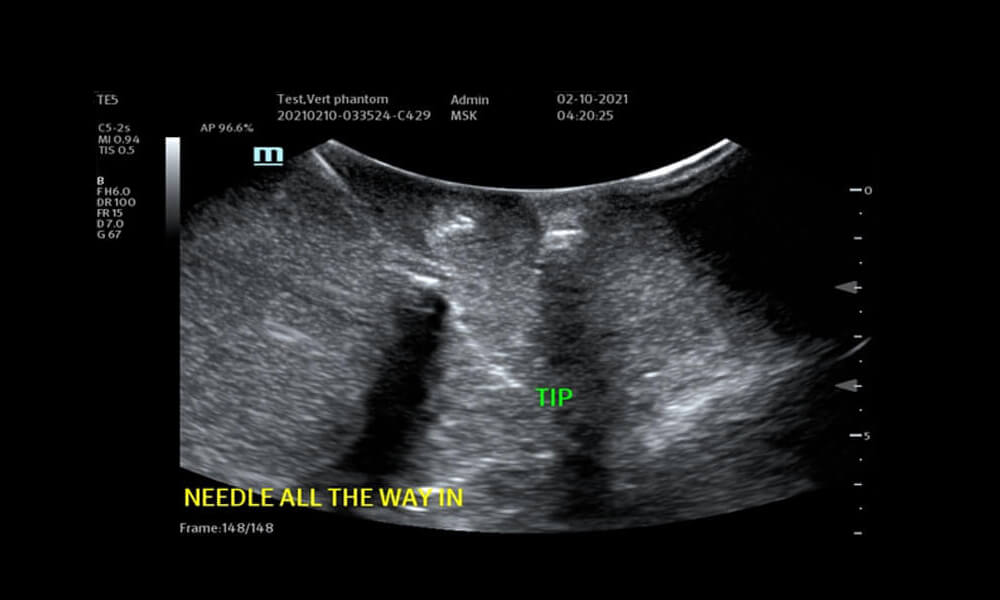
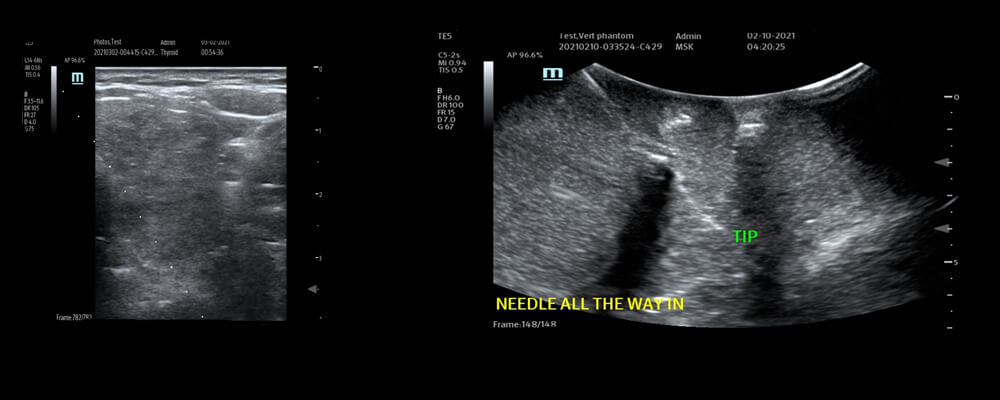
The first one is needle guidance. The science goes by several different names depending on what manufacturer you prefer. It can be performed in 2D, 3D or 4D. This technology cuts across the spectra of sonographic specialities. There are different ways this science is applied. Most commonly it is an algorithm applied to either the needle, the images or both. The most pressing issue when doing any type of needle procedure is seeing where the tip of the needle is in relation to the target. There are several ways to do this, lasers that attach to the transducer, special software like eSpacial Navigation (magnetization), biopsy guides, volume navigation and echo ready needles. Biopsy guides require an attachment to the probe and software to function. This blog will talk about the easiest of all which is iNeedle. This is an algorithm that reads the needle and the tissue. This will track the angle of the needle and show the needle tip beautifully. Unlike a biopsy guide it will also provide a perpendicular purple area so you can have an extra visual guide to placement. The purple area moves with the angle of the needle tip. We tested this extensively and found that it will track angle needles from 25 degrees to 54 degrees. While the variance in angle is great you have to remember that the inline side to side position of the probe has to be straight with the shaft of the needle to work best. The slice thickness is about the size of a side of a credit card.
Please see the pictures below.
© Lisa Bachan for Ultra Select Medical 2021
You can see that without the tip label the needle tip and shaft are hard to see in the above image
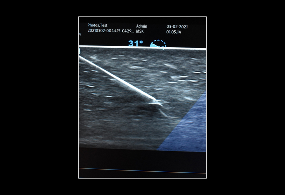
Photo Credit Lisa Bachan
You can clearly see the shaft, needle tip and the degree level that the needle is entering the tissue. There are no brackets or other objects attached to the transducer. No other equipment is needed. This is an 18 gauge needle. It is very simple to use with no limitations. Anyone who needs to do a needle stick will love this.
The next must have is automatic follicle measurements. Some call it Smart FLC; some call it 5G Follicle or Sono AVC. This is a heaven sent feature for anyone in the infertility space. This will automatically measure and stratify many follicles all in one 3D volume rendered image. The follicles are color coded and numbered for easy identification. The important thing is that you must have a 3D/4D transvaginal probe and the option in the ultrasound machine has to be turned on or permanently installed. In the volume rendered image you can move the array of follicles in many directions to have a detailed look at specific follicles.
Each manufacturer has their own cut off as to what is a valid follicle and the measurement applied to them.
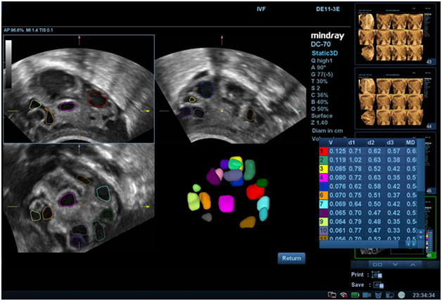
Photo Courtesy of Mindray
With this technology you can delete measurements that you feel is not accurate. You can also merge two adjacent follicles if you need to. Then the measurement matrix can be added to the structured report. This technology increases the efficiency of the ultrasound exam time. With potentially 30 follicles on each side this will be invaluable.
The third technology must have is Elastography. Elastography uses low frequency sound waves to assess the stiffness and elasticity of organs. Currently in the United States elastography has been approved for liver, thyroid, breast and prostate. Other uses will be approved by the FDA when there are nationally normed references for each organ and the research to support the normals. There are two basic types of elastography in our area of ultrasound. The first is strain elastography. This type involves having the patient be still and holding the probe very still. Using consistent manual pressure on the organ is also a technical challenge. This type may work well for superficial organs but not as well for deeper structures.
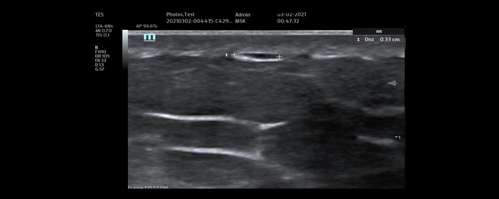
Photo Credit Lisa Bachan 2021
The second basic type is Shear Wave Elastography. SWE uses the transmitted vibrations into the tissues to discern whether or not there is pathology and how stiff it may be. This helps guide treatment if you can have better information about the organ in question. There are several indices that now quantify areas of pathology. When fully approved in all areas including veterinary medicine this will be invaluable. Again it will take the proper training to have the scanning techniques right to make a good diagnosis. We will be offering this training. Stay tuned for future articles under research or clinical trials for new updates on this.
This is a patient who had a COVID vaccine three days prior and still experienced pain in the left lateral deltoid. This the first image to show that there was still fluid in the site. There was no abnormal color flow or Power Doppler. Then it was imaged again on a Mindray DC 88 to be examined with elastography.
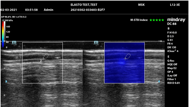
Photo credit Lisa Bachan
As a baseline Shear Wave Elastography (SWE) was used to check the normal skeletal muscle. You can see that the blue shades are consistent and there are five green stars to tell you the confidence level in the image. There are numbers applied in the area of the trace. Now that this was documented then we looked at the area of the COVID injection site.
Even though you cannot see the same small fluid collection that you could on the above image you can see there is an area of stiffness in the green yellow area. The green stars still indicate a good image and that it was done properly. Just based on the image with no numbers you can see that there is increased stiffness. The closer you are to the red the stiffer the object is. Now look at the 2D image to the left. It looks completely normal. The patient was advised to take anti inflammatory and use a warm compress.
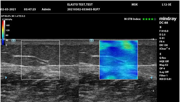
Photo Credit Lisa Bachan
The fourth great technology is Auto EF. This term is basically standard. It refers to the automatic ejection fraction measurement of heart structures. Most sites apply it to the left ventricle. Until recently the EF was obtained by direct visualization (eyeballing it), bi plane 2D or m-mode measurements or 3D measurements. To be truly accurate in the ejection assessment you need several measurements in different segments of the heart muscle. This can be time consuming. So auto EF was born to see border detection with tissue differentiation and plot the points around several segments in the heart structures.
Then the numbers that are generated give a more global ejection fraction. This is based on artificial intelligence and algorithms. This is not the same as strain imaging of the heart. That will be the subject of another article. Some manufacturers have it as an option and others do not. It is worth asking that question prior to any purchase.
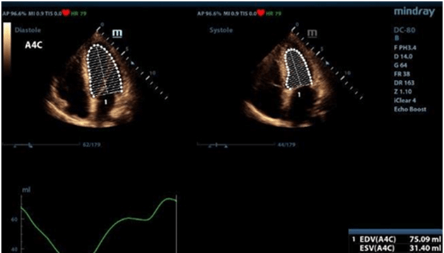
Photo Credit Mindray DC 80-A.
Here you can see two images, one in diastole and one is systole. The results are in volumes, CO and EF all at once. You can see that there are several segments of the chamber walls that are measured. Image quality and operator skill is still necessary for a good outcome. This is truly a time saver and more information is provided.
This makes accuracy and scanning a challenge. Like the others mentioned above this was born from a need to try to whittle down the scanning time and efforts of the operators. This one refers to the head measurements; BPD, OFD, HC, Cerebellum and any IT measurements that may be incorporated into each package. Mindray has it separated into Smart CNS Planes and Smart OB. Others separate out just for the IT measurements. You need to start out with a good 3D of the head at the Cavum Septum Pellucidi near the Thalamus. Then in one good 3D sweep you have all the head measurements. They all go directly to the structured report. Again you need crystal clear structure visualization on 2D in order to get good 3D. Increased accuracy and time savings make this one a must have.
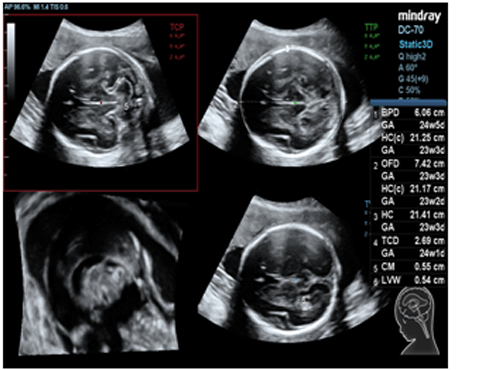
Photo courtesy of Mindray DC70
These great innovations will never replace the skills necessary for great imaging. Nor will they ever solve the issues with obese patients but they will make life easier. Stay tuned as we evaluate the new machines with new features. If you have questions please please contact us at 843-566-2010.