
Structured reports were born in the 1980’s. They were very rudimentary and the computer technology was not at the level to support the true form of the desired outcome. Fast forward to the 2020’s and there is a great deal we can do with structured reports now. Today it is a serious issue if the ultrasound machine and the PAC’s system cannot support structured reporting.
Why were they developed to begin with? The answer was to provide a uniform, easier and faster way for the reading physicians to produce the final reports that will drop into the patient’s chart. Today that process is done electronically and the charts are now subsections of an Electronic Medical Record (EMR). The other reason they were implemented was to standardize the lexicon for each specialty. This approach spans neurosurgery to Ankle Brachial Index (ABI). Ultrasound has a particular language that we use to convey our findings and data in a uniform manner. We need this uniformity because our reimbursement is so closely tied to the coding.
How do we do this? There is programming built into the ultrasound machines that tell the ultrasound machines what formats to send the images and data in. Then the PAC’s or PC has to have the ability and or software to receive the studies and convert it into a format that the reading physician can read and enter in the interpretation and other information to produce the final report. In the past these reports were dictated by the doctor and transcribed by the transcription department. Then these reports were reviewed, signed and dated by the reading physician. They were placed in a paper chart or scanned into the electronic chart. Structured reports strive to simplify this process. If you can take a Structured report file from the ultrasound machine and all the doctor has to do is select from a list of pre made interpretations, sign and date it the with a push of a button it goes into the chart then the process is much more efficient.
What is the manner in which it goes from the ultrasound machine to the PACs or PC? If we talk about structured reports then we must talk about what DICOM is. This format used to transfer the studies from the Ultrasound machine to the PACs. DICOM means Digital Imaging and Communications in Medicine. It came into being from the minds at the National Electronics Manufacturers Association and the American College of Radiology: to have a standardized method of transmission to the permanent storage device. Patient’s studies are never to be stored for long periods of time on the ultrasound machines. Therefore they have to live somewhere. Their final home is either a server, PACs or a PC. Because all of these permanent storage devices are different there had to be a standardized way to transmit the patient studies. There also had to be a way to manage workflows as well as queries. Another issue was having the same device talk to other devices by different manufacturers. DICOM solves all those issues. When ultrasound manufacturers develop their systems, they have to build them with DICOM conformance and compatibility and observe all those protocols. Likewise with the PACs, they have to be built with DICOM in mind. If you are using a PC then you need a DICOM viewer to see the images and reports
What are PACs/DICOM servers and how do they work? DICOM servers – much like viewers are specialized programs that implement the features from the DICOM protocol. A DICOM server can store, query, and retrieve DICOM files over a network – granted the client and server machines are networked and the applications are configured correctly. Instead of simply viewing DICOM files locally, a DICOM server facilitates a centralized storage place for various patient exams. These exams are typically sent from various machines across the network. Any image can be encoded in a DICOM format (.dcm file extension), including structured reports. Most DICOM software will also have options to “export" DICOM images as other formats (JPEG, PNG for images or PDF in the case of structured reports).
What are the differences between PACs and DICOM servers? PACs and DICOM are not one in the same, although they are very closely intertwined and share much of the same functionality. PACs provide storage and convenient access to medical images such as ultrasounds, MRIs, CTs, and x-rays. … PACS use digital imaging and communications in medicine (DICOM) to store and transmit images
DICOM is both a protocol for transmitting images and a file format for storing them, but it ends at the “archival" layer. This leaves PACs vendors with the responsibility of actually storing images. PACs vendors have addressed this difficulty of migrating data from systems and have developed
a more “Vendor neutral" attitude towards data stored in their systems. The acronym “VNA" (Vendor neutral archive) often appears to describe interoperability between PACs. More info can be found in this article: https://www.ncbi.nlm.nih.gov/pmc/articles/PMC3698882/
What happens if my machine does not have structured reports? Most machines today have the ability to send structured reports. There are still some older machines that do not have SR for all body parts. In these cases the PACs will not “see” the headings and measurements. The reports remain “unseen” and the measurements are not seen. The numbers need to be placed in specific bins in the PACs structured report to be seen. What happens if the ultrasound machine cannot be altered to configure it correctly? Then there is no other option but for the PACs company to map each measurement individually for each body part. Sometimes the issue lies between a single word. The PACs may see the word average as such but the ultrasound machine calls it a mean and vice versa. Again in this instance the PACs Company needs to map the term. Mapping is a long and labor intensive process. Every ultrasound manufacturer includes a DICOM conformance statement with each model offered. This can be used by programmers to properly map the SR.
Below are three ways to see information.
1. The worksheet- This is what the sonographer sees when they push the report or worksheet button on the ultrasound machine. This is where we can add, delete and type comments in under certain headings.
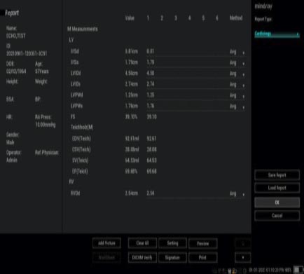
Many times these worksheets are not saved as an image. If they are saved then it is only as an image not as another type of file. Then select a setting that says report, print view or preview to bring you to the PDF report.
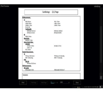
2. PDF. This is what the PAC’s or PC sees when you export a report with your images. This is what is used to make the structured report. This particular one is page 2 of 3 pages of the report. Notice how the headings and numbers are placed on the page. The SR report has to know where to look for it.
3. This is what an actual structured report really looks like. The titles of the measurements are very different. The actual ultrasound images come over in a library that may require the doctor to click on each one or if the PACs or PC is advanced enough they will be seen as a grid. The headings are different as well.
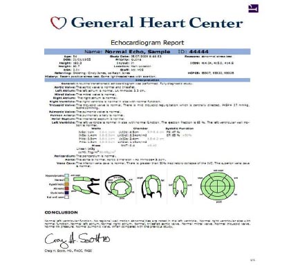
4. The below is the final report that goes into the EMR. So you can see that there are very different functions and appearances of each.
Structured reports are they friend or foe? From the management perspective they are a time saver and a standardization tool. From an ultrasound connectivity version they can be challenging. The connectivity and mapping of the structure isn’t always the same on each end. We get calls all the time saying the structured reporting does not work because the reports do not go over to the PACS or PC. 99.8% of the time it is the PACs that has not been configured properly. The fact is that the DICOM standard is very old at this point. Ultrasound technology is constantly changing and constantly being perfected. Ultrasound manufacturers and PACS developers have to meet these new challenges using existing DICOM technology In the future it will be faster and more AI will be used to have automatic coding and reporting.
Please call us with any questions 843-566-1020.