
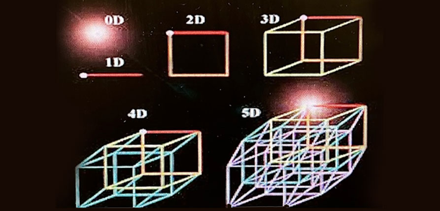
This blog comes to you as a result of a customer request. This is our depiction of the dimensions used in ultrasound. There is a relationship between the math and the images we produce on the monitor. We will also correct some misconceptions along the way.
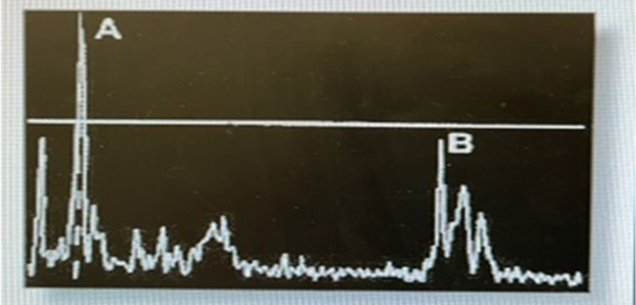
Before we had actual images that resemble the organs, we had the A dimension or A Mode. This was a single line produced by a single ultrasound beam and plotted on a graph in relationship to depth. The evaluation of the eye was the primary use of this type of sound wave. Then it was used in the urinary bladder without much success. Today it is used in focused ultrasound but the results look very different and the application to cancer and brain treatment is much more sophisticated.
Then came 2D (B for brightness mode) ultrasound. Now there is an image showing relationships to other organs. The first generation of this type used the articulated arm holding the transducer. Then you had to divide up the abdomen into a grid that was used by surgeons. This is where we get the umb +1 or -1, ML to left is _1, 2, 3cm etc. To make the image darker you had to retrace the single sweep in the exact same way you did the first sweep. Doing twenty exams a day was not a possibility.
Today we use the terms 2D or B mode interchangeably. There is another term for how we scan today and it is real time. Our images are moving according to the motion of our hand holding the transducer. Pixels are imaged in two dimensions of length and height or width and height depending on your projection. Your measurements are performed in the Length x Width x Height format. These images are in shades of gray and this is also called grayscale imaging. Pixels were the smallest unit of dot that was picked up by our machines. They are assembled into images at an extremely fast speed.
Fast forward 20 years and we have static 3D imaging. The primary use of these images is in OB/Gyn. There are other applications in vascular, pediatrics and cardiac but the majority of users are in OB. Now all three dimensions of length, width and height are compiled from three planes (A, B and C) into a volume rendered image (VR). Each plane can be rotated on three axes X, Y and Z to optimize the VR image. Instead of pixels 3D uses Voxels. The color of the VR image is not an indication of a level of dimensionality. The color is simply a tint map or other algorithm applied to the original gray image.
What is noticeable to anyone who has scanned in 3D is that if you were to turn the above image around, the back of the head and neck would not have a VR image. It is just the front part of the face and in this case the hand that is rendered. As long as the measurements are applied to the completed VR image you can get three measurements. You could possibly take the 2D measurements on each plane except the VR image and still have all three measurements in all three dimensions.
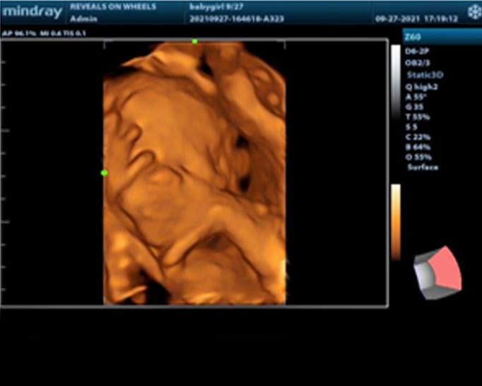
We have the answer to the above problem and it is 4D ultrasound. 4D is 3D but in motion. 4D is always imaged by using cine clips unless you freeze the clip and then save it as a still image. If the above image was done in 4D you could turn it around and get a fast rendering of the back of the baby’s head and neck thus imaging in the 4th dimension. This dimension is what is in back of the initial image and being able to see it in a VR image. This is also called whole rendering.
There are a vast array of changes you can make to the 3D and 4D images. You can change light sources, depth tints, some smart functions can delete overlying structures. Plus you can physically cut out unwanted areas with an edit tool. None of these tools change any 3D or 4D image into 5D.
There are advertisements that say 5D and 5G in ultrasound. This is not correct. First let us tell you what the fifth dimension really is and why it is not in any ultrasound machine. We had someone call us and tell us that if the image is not pink it is not 5D. Hmmmm. This is not correct either. Colors never dictate dimension.
The fifth dimension is not visible. If you cannot see it you cannot measure it. This has been a mantra in ultrasound since ultrasound was invented. It is considered to be a micro dimension because of how this would have to be linked to the Huygens Principle. 5G refers to the generation of cell phone technology ONLY. There have been experiments in England and China using 5G in conjunction with 3D and 4D ultrasound , using a robotic hand to scan. The premise is that with 5G you can scan remotely and image real time in emergency situations.
The other reason that ultrasound cannot image in the 5th dimension is that mathematically it is nearly impossible to create a formula to relate the 5th
dimension to the Huygen’s principle. Ultrasound machines cannot handle the use of the Kaluza-Klein math or allow a . These integrals and multilevel mathematics are simply too large for ultrasound machines. It would require some alteration of a Petrov III time – space continuums. “There exist no Petrov type III space- times on which Maxwell equations or Weyl neutrino equations satisfy Huygens Principle.” Physicists have been trying for centuries to crack the 5th dimension.
Just keep in mind that features like HD Live, HD Silhouette, iLive and what one manufacturer calls 5G will not allow you to truly be in the 5th dimension. They are just tools that allow you to change a light source, image with a more realistic skin tone or remove structures. Just be thankful we do not have to learn 5D in physics.
Reference
Huygens’ Principle for Relativistic Wave Equations on Petrov Type III Space-Times. Author Fernando Deeke Sasse 1997. Doctoral Thesis in Philosophy and Applied Mathematics. University of Waterloo. Canada